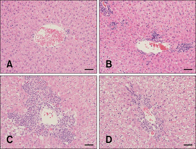
Fig. 2. Histological features of liver sections. APAP-treated rats exhibited minimal to moderate centrilobular single-cell necrosis and/or apoptosis with inflammatory cell infiltration. (A) In the 6 h group, the histology of the liver was normal. (B) In the 24 h group, minimal to slight centrilobular necrosis was observed. (C) In the 48 h group, moderate to marked centrilobular necrosis with inflammation were observed. (D) In the 72 h group, slight to moderate centrilobular necrosis was observed. H&E stain. 200× (A–D). Scale bars = 50 µm.

