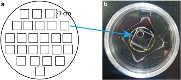
Fig. 3.

PDMS stencil for cell patterning. (a) Template printout for trace-cut cured PDMS in a 100 mm petri dish. (b) Each PDMS square dimension is 1 cm by 1 cm to fit into the glass well, indicated by yellow dash line, of a 14 mm glass-bottom dish. A circle with Ø=0.4 cm is punctured through the PDMS square to create the stencil space for cFB cluster
