
Fig. 5.
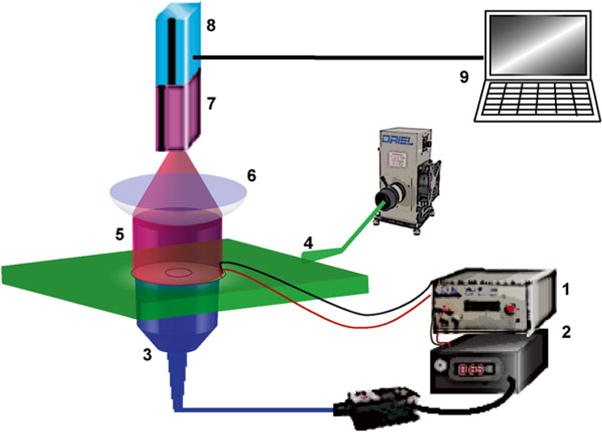
Ultrahigh-resolution high-speed optical imaging system was used to capture electrically and optically induced excitation waves, reported by calcium-sensitive fluorescent dye Rhod-4 or voltage-sensitive fluorescent dye FluoVolt™: (1) Pulse generator for electrical pacing (analog modulation) and optical pacing (TTL modulation). (2) Fiber optic-coupled 470 nm laser as activation light source for ChR2 or 590 nm LED as activation light source for ArchT. (3) Global illumination of co-cultured ChR2-cFB/cardiomyocytes or ArchT-cFB/cardiomyocytes was delivered from below the sample. (4) Excitation for Rhod-4 or FluoVolt was delivered to the sample as a light sheet from a filtered arc lamp. (5 and 6) Rhod-4 and FluoVolt emission fluorescence was collected through appropriate filters for high-resolution macroscopic lens. (7) Gen III MCP intensifier. (8) Pco 1200hs CMOS camera. (9) Computer system and software for image acquisition