
Fig 2.
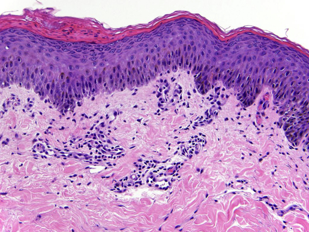
Patient 3. Biopsy specimen from the left inguinal area shows slight spongiosis and superficial perivascular infiltrate with occasional neutrophils and focal basal layer hydropic degeneration. (Hematoxylin-eosin stain; original magnification: ×40.)