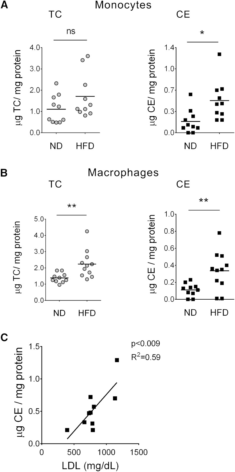
Fig. 1.
Peripheral blood monocytes and peritoneal macrophages from hypercholesterolemic mice accumulate CE. A, B: Total cholesterol (TC) and CE in peripheral blood monocytes (A) and peritoneal macrophages (B) isolated from LDLR−/− mice fed a high-fat diet (HFD) or a normal diet (ND). TC and CE levels were normalized to cellular protein content. Each point represents an individual; horizontal lines indicate mean, n = 10/group. ND versus HFD, * P < 0.05, ** P < 0.01, ns = nonsignificant (t-test). C: Positive correlation between CE content in circulating monocytes from HFD mice and the plasma LDL. Each point represents an individual, n = 10 (Pearson correlation, P < 0.009; R2 = 0.59).

