FIG. 4.
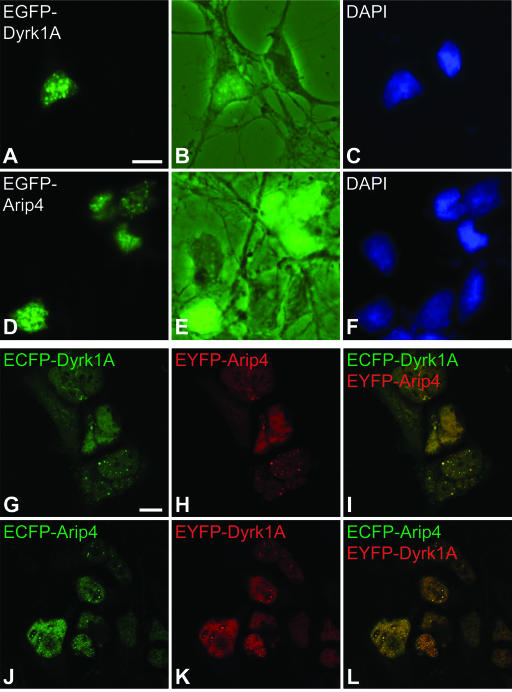
Intracellular localization of Dyrk1A and Arip4 in primary hippocampal neurons and in HEK293 cells. (A to F) Primary rat hippocampal neurons were transfected with EGFP-Dyrk1A (A to C) and EGFP-Arip4 (D to F). (A and D) Fusion proteins were detected by conventional fluorescence microscopy. (B and E) To visualize entire cells, corresponding images were taken with phase-contrast settings in combination with fluorescence. (C and F) Nuclei were counterstained with DAPI. Note that both EGFP-Dyrk1A and EGFP-Arip4 were localized in a speckle-like nuclear subcompartment. (G to L) Fusions of ECFP and EYFP with Dyrk1A and Arip4 were coexpressed in HEK293 cells and were analyzed by confocal laser scanning microscopy. ECFP signals (green; G and J) and EYFP signals (red; H and K) were merged (I and L) to visualize colocalization (yellow). Scale bars: A to F, 5 μm (bar shown only in panel A); G to L, 10 μm (bar shown only in panel G).

