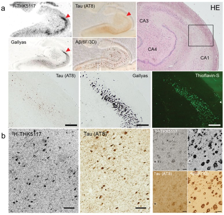
Figure 3.
(a) In vitro autoradiography of 18F-THK5117 and tau/amyloid immunohistochemistry, H&E staining, Gallyas silver staining, and thioflavin-S fluorescence staining in the hippocampus of AD brain sections. Scale bars: 400 μm. (b) Microscopic observation of 3H-THK5117 and 3H-PiB labeled sections after photo emulsion treatment and tau/amyloid immunohistochemistry in adjacent sections. (asterisks indicate same blood vessel, scale bars 100 μm).

