FIG. 4.
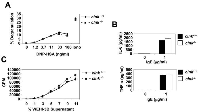
Analyses of mast cell functions. (A) Antigen-induced degranulation. BMMCs were sensitized with anti-DNP IgE and triggered by addition of the indicated concentrations of the antigen DNP-HSA. Degranulation was assessed by measuring the release of β-hexosaminidase in the supernatant. Control degranulation was examined in the presence of ionomycin (Iono). Values are presented as percentages of maximal release. Degranulation was measured in response to DNP-HSA. (B) IgE-induced cytokine secretion. Cells were stimulated for 3 h with the indicated concentration of IgE, and secretions of IL-6 (top panel) and TNF-α (bottom panel) were determined by ELISA. Similar results were obtained over a range of different IgE concentrations (data not shown). Standard deviations are represented. (C) IL-3-induced proliferation. BMMCs were incubated in the absence of IL-3-containing medium for 8 h. Then, they were cultured for 18 h in the presence of the indicated concentrations (represented as a percentage of final volume) of WEHI-3B supernatant (a source of IL-3). Proliferation was determined by measuring thymidine incorporation. Standard deviations are represented.

