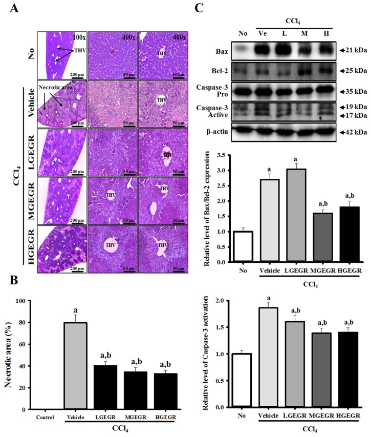
Figure 3.
Histology of liver and expression of apoptosis-related proteins. (A) Liver tissues collected from ICR mice were fixed in 4% formalin and stained with H&E solution. Liver tissue was mainly observed around the terminal hepatic venule (THV) at a magnification 200× and 400×; (B) The necrotic area in each section was observed using Leica Application Suite (Leica Microsystems, Switzerland); (C) Expression levels of caspase-3, Bcl-2 and Bax proteins in the liver tissue were analyzed by western blot analysis. Membranes were incubated with antibodies for caspase-3, Bcl-2, and Bax, as well as β-actin protein from the liver. Expression levels were quantified by an imaging densitometer, and the sizes of the products indicated. Data represent the means ± SD of three replicates. a, p < 0.05 compared to the no treatment group; b, p < 0.05 compared to the vehicle/CCl4 treated group.

