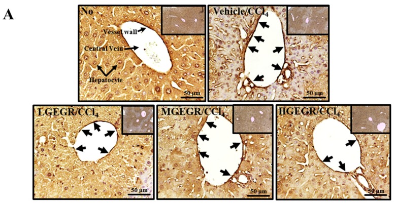
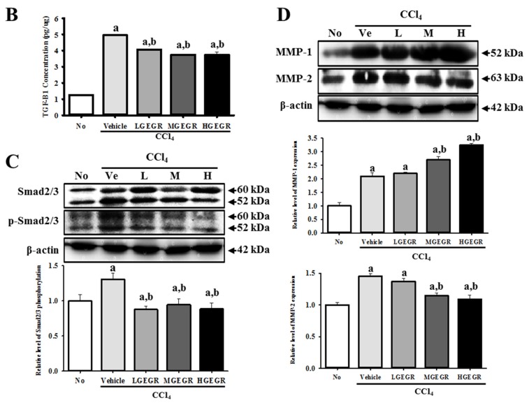
Figure 6.
Analysis of the regulatory factors of liver fibrosis. (A) Immunohistochemistry of collagen protein. After GEGR/CCl4 pretreatment for 5 days, the liver tissues were collected from all groups and slide sections of liver were prepared as described in the Materials and Methods. The distribution of collagen protein in the slide sections of liver tissue was determined by staining with collagen specific antibody followed by observation at 400×. Black arrows indicate the stained area in THV; (B) ELISA for TGF-β1. After collection of blood serum, the concentration of TGF-β1 was determined in the serum collected from the brains of mice using a TGF-β1 ELISA kit that could detect TGF-β1 at 3.5 pg/mL; (C) Western blot analysis for Smad2/3 expression. To measure the expression levels of Smad2/3 and p-Smad2/3 protein, the membranes were incubated with specific antibodies for each protein, as well as β-actin protein from liver lysates. Three mice per group were assayed by western blotting; (D) Western blot analysis of MMP expression. To measure the expression levels of MMP-1 and MMP-2 protein, the membranes were incubated with specific antibodies for each protein, as well as β-actin protein from liver lysates. Three mice per group were assayed by western blotting. Values are reported as the means ± SD. a, p < 0.05 compared to the no treatment group; b, p < 0.05 compared to the vehicle/CCl4 treated group.


