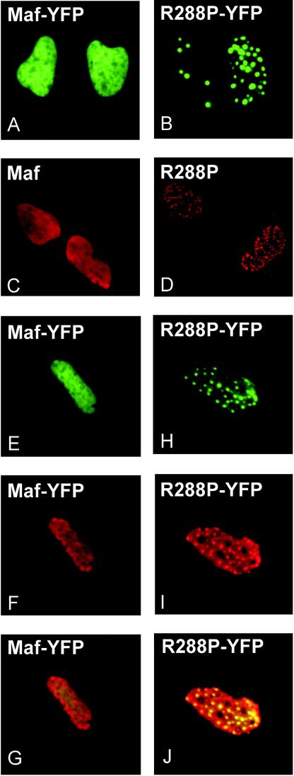
FIG. 8.
Effect of the R288P mutation on Maf localization. Plasmids encoding the proteins indicated in each panel were transfected into αTN4 cells. The distributions of the proteins were visualized by YFP fluorescence (A, B, E, and H) or immunolabeling with Maf-specific antibody (C, D, F, and I). Panels G and J show the overlay of the YFP fluorescence and the immunoreactivity, confirming that the immunoreactivity reflected the localization of the proteins.

