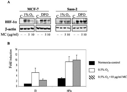
FIG. 4.
(A) The effect of MC treatment on HIF-1α levels in MCF-7 and Saos-2 cells. Cells were seeded at 40,000/cm2 and treated as described in Fig. 1A. Total protein lysates (40 μg) were tested for HIF-1α and β-actin by Western blotting. (B) Cotransfected HIF-1α rescues CA9 promoter activation in MC-treated MCF-7 cells. Cells were cotransfected with the [−173, +31] CA9 reporter construct, pRL-tk, and 500 ng of pCEP4 (EV) or pCEP-HIF-1α (HIF-1α) as in Fig. 2. CA9 promoter activity in the presence of EV in control cells (21% O2) was set as 1, and the rest is expressed as the level of induction as in Fig. 2.

