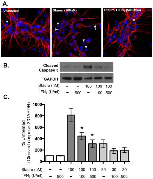
Figure 5. IFNγ blocks apoptotic signaling in neurons upon staurosporine exposure.
Cultured hippocampal neurons were treated with IFNγ at the indicated concentrations (100 or 500 U/ml). After 30 min of pretreatment with IFNγ, staurosporine (30 or 100nM) was added to the cultures for 72 h. A. Cells were fixed and stained for MAP2 (red) as a marker for neurons and Hoechst (blue) for cell nuclei. Arrowheads indicate pyknotic nuclei and arrows indicate beaded dendrites. B. Cells were lysed in protein solubilization buffer and analyzed by Western blot with antibodies against cleaved caspase 3 and GAPDH as a loading control. C. Western blots were quantified by densitometry using ImageJ software. Cleaved caspase 3 signal was normalized to GAPDH as a loading control and statistical analysis was determined using a paired t test (n = 4, *p < 0.04 versus signal in untreated control).

