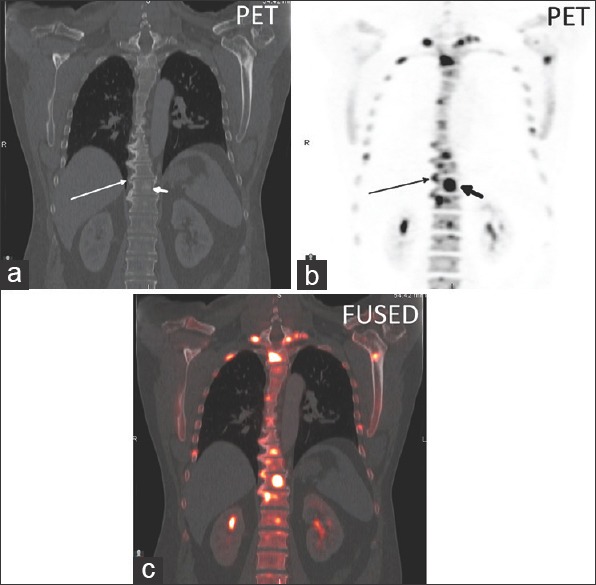
Figure 2.

(a) Coronal CT, (b) coronal 18F NaF PET, and (c) axial fused PET/CT image in a thoracic spine degenerative disc osteophyte (long arrow) with a SUVmax of 27.3 and an adjacent sclerotic vertebral body metastasis (short arrow) with a SUVmax of 64.0
