FIGURE 1.
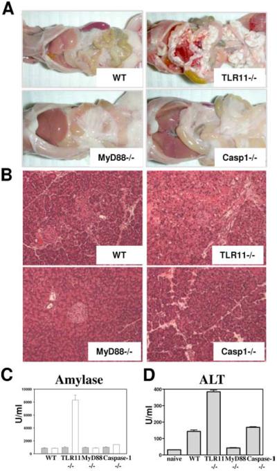
TLR11-deficient mice develop acute pancreatitis in response to T. gondii infection. A, Development of fat necrosis in T. gondii-infected TLR11−/− mice. WT, TLR11−/−, MyD88−/−, or Caspase1−/− were infected with an average of 20 cysts per mouse of the ME49 strain of T. gondii and the peritoneal cavities were examined 5 days later. B, Animals were infected as described above and pancreatic tissues were removed for histological analysis (H&E staining). The images shown are representative of multiple sections examined in three or four mice per group. C, WT, TLR11−/−, MyD88−/−, or Caspase1−/− were infected as described above and 5 days later serum levels of amylase were analyzed; □, T. gondii infected;  , naive controls. The data are representative of eight experiments performed. D, Serum levels of inflammation marker alanine amino-transferase (ALT) in mice infected with T. gondii. The data are representative of three experiments performed.

