
Fig 2.
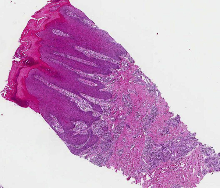
Skin biopsy specimen from a skin lesion showed hyperkeratosis, papillomatosis, and acanthosis with elongation of the rete ridges and perivascular inflammatory cells in the dermis. (Hematoxylin-eosin stain; original magnification: ×40.)
Official websites use .gov
A
.gov website belongs to an official
government organization in the United States.
Secure .gov websites use HTTPS
A lock (
) or https:// means you've safely
connected to the .gov website. Share sensitive
information only on official, secure websites.
Skin biopsy specimen from a skin lesion showed hyperkeratosis, papillomatosis, and acanthosis with elongation of the rete ridges and perivascular inflammatory cells in the dermis. (Hematoxylin-eosin stain; original magnification: ×40.)