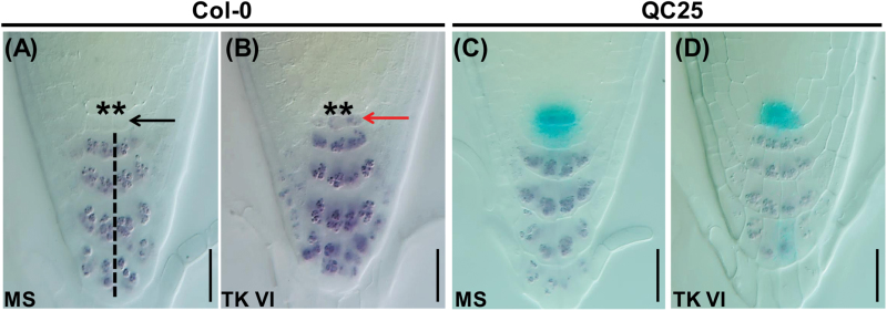
Fig. 3.
TK VI-induced disruption of the maintenance of the root stem cell niche in Col-0.
(A, B) TK VI-induced CSC differentiation in Col-0 as shown by Lugol staining. Six-day-old seedlings were transferred to medium without (MS) (A) or with (B) 5 μM TK VI for 12h before Lugol staining (dark brown). The asterisks denote QC cells. The black dashed line indicates the columella cell layers. The black arrowhead indicates a lack of starch accumulation in non-differentiated CSCs. The red arrowhead shows starch accumulation in TK VI-treated CSCs. (C, D) Disorganized QC cells and differentiated CSCs as shown by Lugol staining of the QC25 marker line. Six-day-old seedlings were transplanted to medium without (MS) (C) or with (D) 5 μM TK VI for 48h before double staining with GUS and Lugol was performed. Scale bars=20 µm (A–D).

