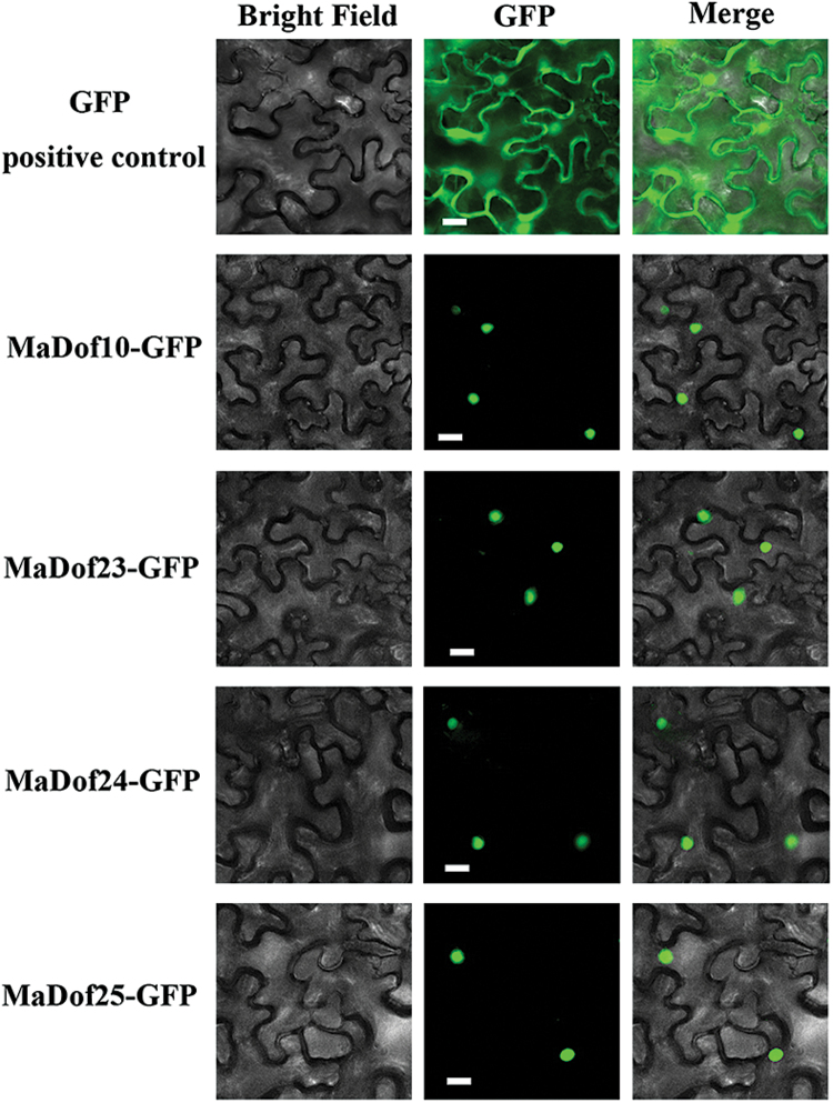
Fig. 3.
Subcellular localization of MaDof10, 23, 24, and 25 in tobacco leaves. MaDof10, 23, 24, and 25 fused with the GFP or GFP positive control were infiltrated into tobacco leaves via A. tumefaciens strain GV3101. After 48h of infiltration, GFP fluorescence signals were visualized using a fluorescence microscope. Merge indicates a digital merge of bright field and fluorescent images. Images were taken in a dark field for green fluorescence, while the outline of the cell and the Merged images were photographed in a bright field. Scale bars, 25 μm.

