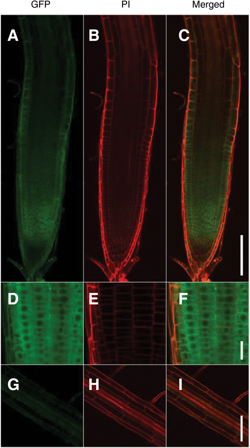
Fig. 6.
Tissue specific expression of TPR5–GFP fusion protein. Three DAG seedlings of transformants expressing TPR5–GFP (genetically identical to tpr5-2 TPR5(genomic)–GFP L1 in Fig. 4) were observed using confocal microscopy. The cell wall was stained with PI. (A–C) Representative images of primary root tips. (D–F) Primary root tips with higher magnification. (G–I) Mature region with root hair. Bars: (A–C, G–I) 100 µm; (D–F) 20 µm.

