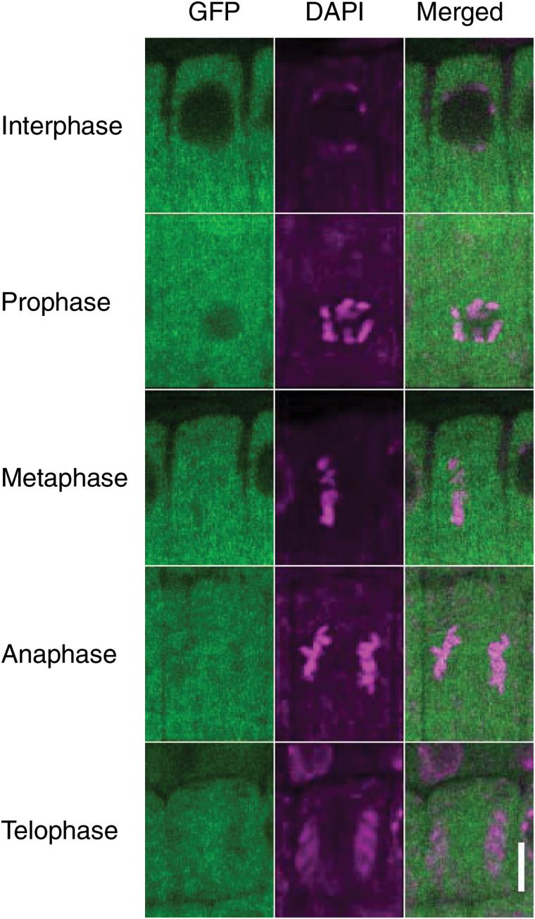
Fig. 7.
Subcellular localization of TPR5–GFP fusion protein during cell division. Three DAG seedlings of transformants expressing TPR5–GFP (genetically identical to tpr5-2 TPR5(genomic)–GFP L1 in Fig. 4) were observed using confocal microscopy. DNA was stained with DAPI and root meristematic epidermal cells in each cell division phase were observed. Bar: 5 µm.

