Fig. 3.
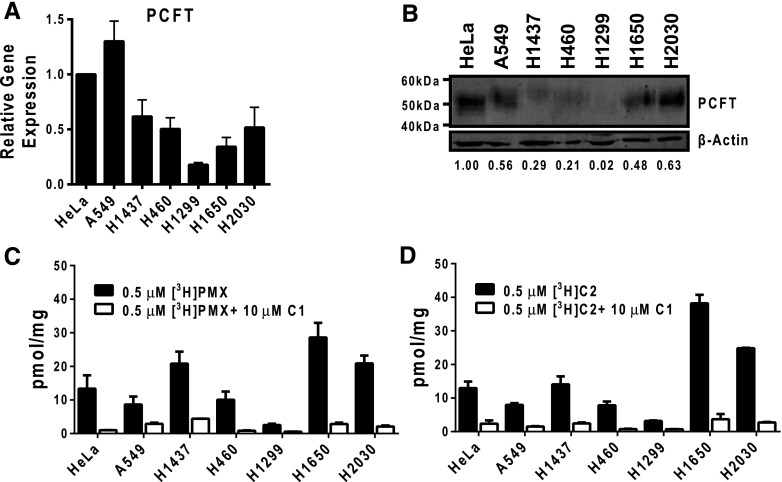
PCFT expression and function in NS-NSCLC cell lines. (A) Results are shown for PCFT transcript levels measured by real-time RT-PCR in NS-NSCLC cell lines (A549, H1437, H460, H1299, H1650, and H2030). PCFT transcript levels were normalized to transcript levels for β-actin. The PCFT transcript level for the HeLa cell line was assigned a value of 1. Results are shown as mean ± S.E. values from triplicate experiments. (B) Particulate membrane fractions were prepared as described in Materials and Methods. Membrane proteins (30 μg) from human tumor cell lines were electrophoresed on a 7.5% polyacrylamide gel and immunoblotted with human PCFT polyclonal antibody. β-Actin levels were used as loading controls. Densitometry was performed using Odyssey software, and PCFT protein expression was normalized to β-actin. Normalized densitometry results (as an average from three experiments) are shown. (C and D) NS-NSCLC cells (in 60-mm dishes) were treated with 0.5 μM [3H]PMX or [3H]C2 at pH 5.5 and 37°C for 5 minutes (black bars). Nonradioactive C1 (10 μM) was added to competitively block PCFT-mediated uptake as a negative control for PCFT transport (white bars). Internalized pmol values of [3H]PMX or [3H]C2 were normalized to total cell proteins and expressed as pmol/mg. Histograms show mean ± S.E. values from triplicate experiments.

