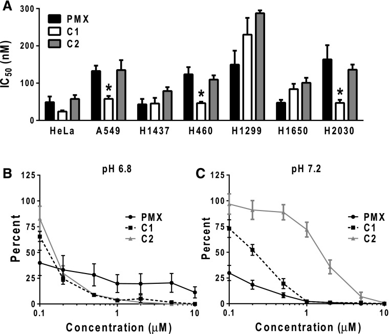
Fig. 4.
In vitro drug efficacy of PMX, C1, and C2 of NS-NSCLC cell lines. (A) NS-NSCLC cells were seeded in 96-well plates at 1500–4000 cells/well in complete folate-free RPMI 1640 (pH ∼7.2), 10% dialyzed fetal bovine serum, and 25 nM leucovorin. Cells were incubated with varying concentrations of PMX, C1, or C2 from 1 to 1000 nM for 4 to 5 days, depending on the cell line. Cell viabilities were determined with a fluorescence-based assay (Cell Titer Blue). Mean IC50 ± S.E. values from triplicate experiments were determined graphically for each drug. Asterisks designate statistically greater sensitivity for C1 compared with PMX (P < 0.01). (B and C) H460 cells (100–150 cells) were plated in 60-mm dishes in complete folate-free RPMI 1640 (pH 7.2), 10% dialyzed fetal bovine serum, and 25 nM leucovorin. After 24 hours, cells were then treated with PMX, C1, or C2 at varying concentrations in complete folate-free RPMI 1640 (at pH 7.2 or 6.8) supplemented with 25 nM leucovorin. After an additional 24 hours, cells were rinsed with PBS, and then incubated in complete folate-free RPMI 1640 supplemented with 25 nM leucovorin (pH 7.2) for 9 days. Colonies were stained with methylene blue and counted, and colony numbers were normalized to the controls. Plots show mean ± S.E. values, representative of triplicate experiments.

