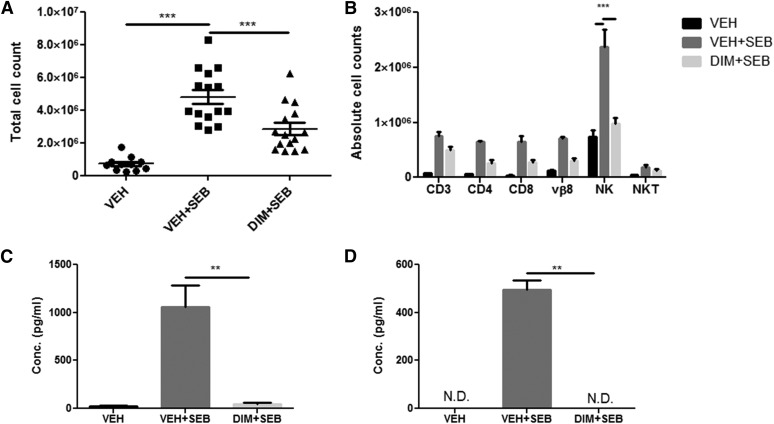
Fig. 2.
DIM decreases immune cell infiltration and IFN-γ expression in the lungs. All experiments were performed 48 hours after SEB exposure. (A) Total number of mononuclear cells from lungs of mice is expressed as per mouse. (B) Immune cells were further stained with monoclonal antibodies to determine the following subsets: T cells (CD3), T-helper cells (CD4), T-cytotoxic cells (CD8), natural killer cells (NK), and natural killer T-cells (NKT). The percentages were multiplied by the total cell number to yield the absolute cell counts shown. (C) IFN-γ levels in BAL fluid. (D) IFN-γ expression in serum. All cytokines were determined using enzyme linked immunosorbent assay (ELISA) from Biolegend. Vertical bars in (B–D) represent mean ± S.E.M. from groups of four or five mice. Analysis of variance, ***P < 0.001; **P < 0.01 with Tukey’s post hoc test.

