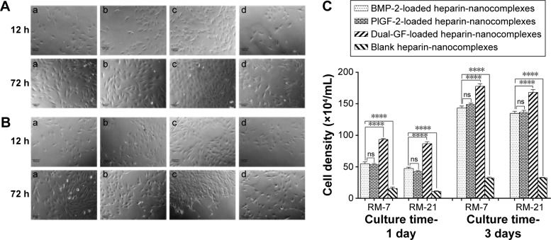
Figure 6.
Microphotograph of MC3T3-E1 cells after 12 and 72 hours of culturing with released medium collected on various nanocomplexes, as observed by using the inverted phase-contrast microscope.
Notes: The released medium was collected on days 7 (A) and 21 (B). (a) BMP-2-loaded heparin-nanocomplexes; (b) PlGF-2-loaded heparin-nanocomplexes; (c) dual-GF-loaded heparin-nanocomplexes; (d) blank heparin-nanocomplexes. The images were obtained at the same magnification (×10). Scale bar =100 μm. (C) Quantitative analysis of MC3T3-E1 cells after 1 and 3 days of culturing with various released mediums. ns>0.05, ****P<0.0001.
Abbreviations: BMP-2, bone morphogenetic protein; PlGF-2, placental growth factor-2; GF, growth factor; RM, released medium; RM-7, released medium collected on the seventh day; RM-21, released medium collected on the 21st day.

