
Figure 1.
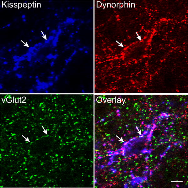
Confocal images (1 μm thickness; 63×) of a section immunolabeled for kisspeptin (blue), dynorphin (red) and vGlut2 (green) in the ARC. White arrows indicate examples of triple-labelled kisspeptin/vGlut2/dynorphin terminals in close apposition to a dual-labeled kisspeptin/dynorphin (KNDy) cell body. Scale bar, 10 μm.