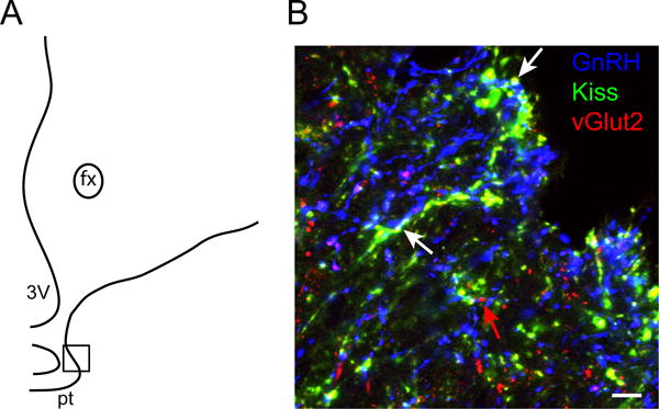
Figure 4.

Dual-labeled kisspeptin- and vGlut2-positive fibers in close association to GnRH fibers in the median eminence. A: Schematic drawing of a section through the ovine median eminence, showing the approximate location of the image (boxed area) shown in B. B: Confocal image (1 μm thickness; 63×) of a section through the ovine median eminence triple-labelled for kisspeptin (green), vGlut2 (red) and GnRH (blue). White arrows indicate examples of dual-labelled (yellow) kisspeptin/vGlut2 fibers and terminals adjacent to GnRH fibers (blue), and the red arrow indicates a single labelled vGlut2 terminal. fx = fornix; pt= pars tuberalis; 3V= third ventricle. Scale bar, 10 μm.
