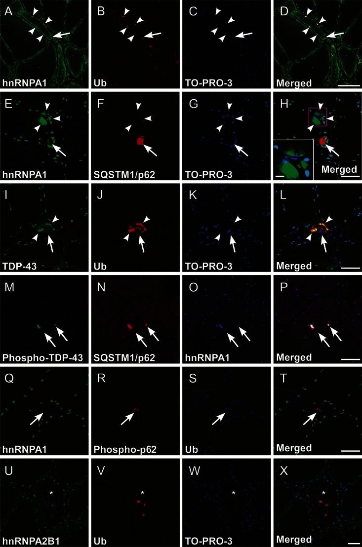
Figure 4. Multiple immunofluorescence for hnRNPA1 and related proteins.
Representative microphotographs of transverse cryosections from the biopsied skeletal muscle of patient III-2 in family 1. (A–H) Aberrant subsarcolemmal/perinuclear aggregation (arrowheads) and increased sarcoplasmic retention of heterogeneous nuclear ribonucleoprotein (hnRNPA1) were mainly evident in atrophic fibers. Inset in H is a higher magnification of the boxed area (scale bar = 10 μm). The subsarcolemmal/perinuclear hnRNPA1 aggregates were often colocalized with ubiquitin (Ub, A–D, arrowheads) and SQSTM1/p62 (E–H, arrows). Note the close association of hnRNPA1/Ub double-positive aggregation with the rimmed vacuole (A–D, arrows). (I–L) In the atrophic fibers, transactive response DNA binding protein 43 kDa (TDP-43)/Ub double-positive aggregation (arrowheads) was also observed with the cytoplasmic mislocalization and nuclear depletion of TDP-43 (arrows). (M–P) The aberrant aggregation of hnRNPA1 was occasionally triple-labeled with phosphorylated TDP-43 and sequestome-1/p62 (SQSTM1/p62), closely adjacent to rimmed vacuoles (arrows). (Q–X) The rimmed vacuoles were often related to Ub, phosphorylated SQSTM1/p62 (Q–T, arrows), and hnRNPA2B1 (U–X, asterisks indicate a rimmed vacuole–carrying fiber). TO-PRO-3: nuclear staining (C, G, K, W). Scale bars = 50 μm.

