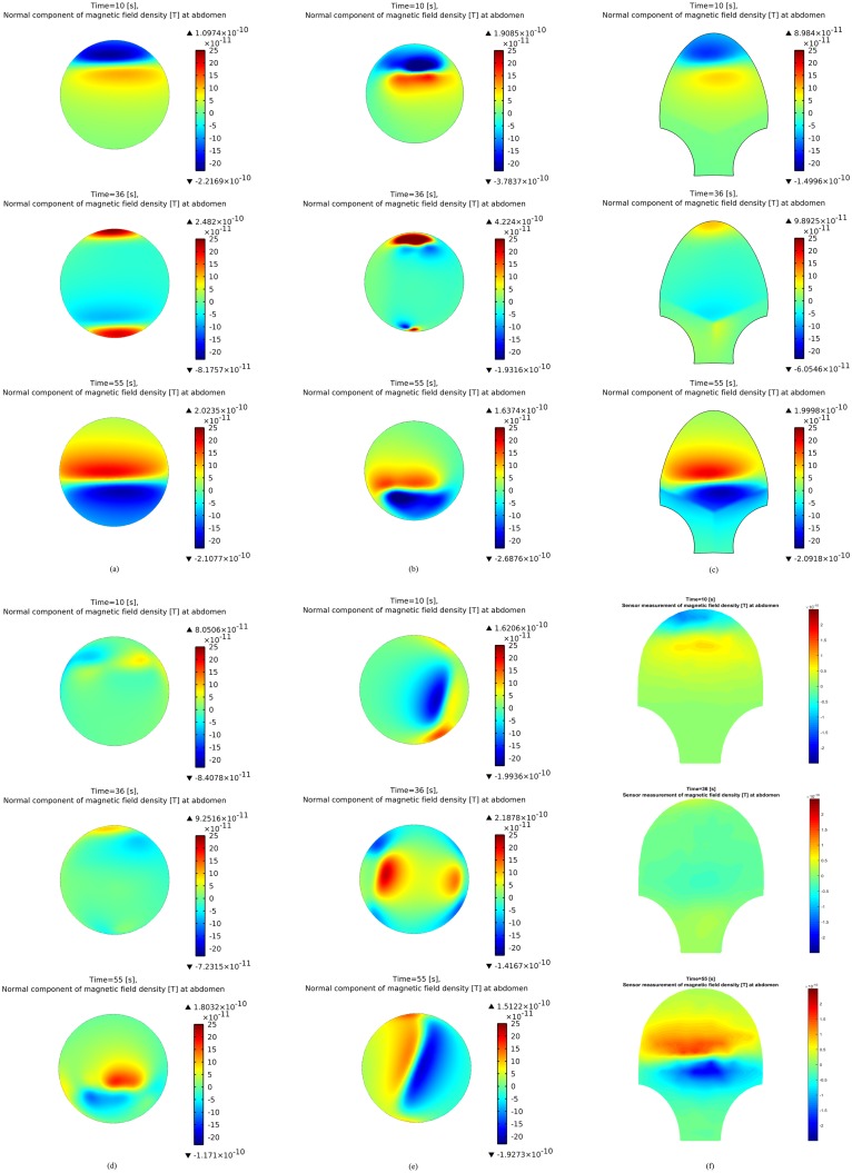
Fig 6. Magnetic field simulations at the abdominal surface at time instants t = 10 s, 36 s, 55 s.
(a) Simulated normal magnetic field with original configuration. (b) Simulated normal magnetic field with anatomical uterus. (c) Simulated normal magnetic field with SARA-shape abdomen. (d) Simulated normal magnetic field with random fiber orientation. (e) Simulated normal magnetic field with pacemaker set at the lateral of uterus. (f) Simulated magnetic field with SARA-shape abdomen and sensor model.

