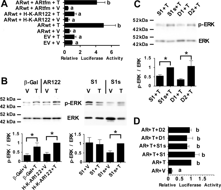
FIG. 3.
H-K-AR122 and S1 are pathway-selective inhibitors of classical or nonclassical T signaling, respectively. A) 15P-1 Sertoli cells transfected with the PSALuc reporter plasmid and plasmids expressing ARwt, ARwt + ARtfm, or ARwt + H-K-AR122 (AR122) were stimulated with vehicle (V) or T for 24 h. The relative luciferase levels were determined relative to empty vector + vehicle (EV + V, =1.0) Error bars show SEM for four experiments. B) Primary rat Sertoli cell cultures were infected with adenovirus constructs expressing β-galactosidase (β-gal), H-K-AR122, or the S1 or control S1s peptide. The cells were stimulated for 15 min with 100 nM T (T) 24 h after adenovirus infection. Western analyses of whole cell extracts are shown for p-ERK and total ERK (T-ERK). Quantitation of p-ERK/ERK values normalized to Ad H-K-AR122 + T (left) or AdS1s+ T (right) (=1) for three experiments is shown below the Western blots. C) Primary Sertoli cell cultures were incubated for 2 h with S1 or S1s peptides (5 μM) or D1 or D2 peptidomimetics (100 nM) prior to a 15-min stimulation with T (100 nM). Western immunoblots for p-ERK and ERK are shown. Quantitations of the p-ERK/ERK values from four experiments were normalized to S1s + T (=1). For B and C, paired one-tailed t-tests revealed statistically significant differences (P < 0.05) where indicated with an asterisk (*). D) The 15P-1 Sertoli cells transfected with the PSALuc reporter plasmid and a plasmid expressing ARwt incubated with S1 or S1s (5 μM) or D1 or D2 (100 nM) during a 24-h stimulation with vehicle (V) or 100 nM T (n = 3). Values with different lowercase letters were found to differ significantly by ANOVA (P < 0.05).

