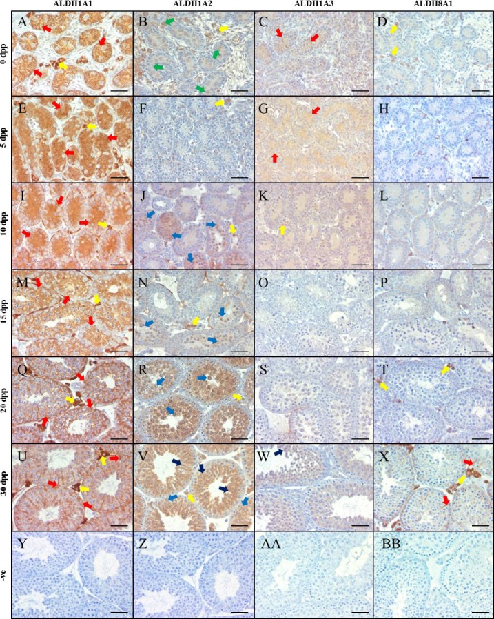
FIG. 1.

ALDH enzymes are differentially localized in the neonatal murine testis. Images are representative murine testicular cross-sections displaying IHC analysis of ALDH localization at various neonatal ages. ALDH1A1 is represented in A, E, I, M, Q, and U for 0, 5, 10, 15, 20, and 30 dpp, respectively. ALDH1A2 is represented in B, F, J, N, R, and V, while ALDH1A3 is represented in C, G, K, O, S, and W for the same time points. Finally, ALDH8A1 is displayed in D, H, L, P, T, and X. Negative controls (−ve) are shown in Y-BB. Brown staining indicates an immunopositive reaction. Arrows indicate immunopositive cells, while the respective colors indicate the following cell types: red, Sertoli cells; yellow, Leydig/interstitial cells; dark blue, spermatid; light blue, spermatocyte; green, gonocyte/spermatogonia. Bars = 100 μm.
