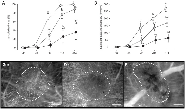
Fig 2. Vascularization of endometriotic lesions in the skinold chamber.
(A, B) Vascularized area (%) (A) and functional microvessel density (cm/cm2) (B) of developing endometriotic lesions in dorsal skinfold chambers of vehicle-treated controls (white circles; n = 6) and 4-MU-treated (20mg/kg: grey circles, n = 8; 80mg/kg: black circles, n = 7) BALB/c mice, as assessed by intravital fluorescence microscopy and computer-assisted image analysis. Means ± SEM. *p<0.05 vs. control; #p<0.05 vs. 20mg/kg 4-MU; ap<0.05 vs. d0 and d3; bp<0.05 vs. d0, d3 and d6; cp<0.05 vs. d0, d3, d6 and d10. (C-E) Intravital fluorescent microscopic images of endometriotic lesions (borders marked by dotted line) at day 10 after transplantation of endometrial fragments into the dorsal skinfold chamber of a vehicle-treated control (C), a 20mg/kg 4-MU-treated (D) and an 80mg/kg 4-MU-treated (E) BALB/c mouse. Blue light epi-illumination with contrast enhancement by 5% FITC-labeled dextran 150,000 i.v. Scale bar: 200μm.

