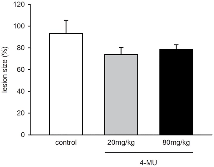
Fig 3. Growth of endometriotic lesions.

Lesion size (%) of endometriotic lesions at day 14 after transplantation of endometrial fragments into the dorsal skinfold chambers of vehicle-treated controls (white bar; n = 6) and 4-MU-treated (20mg/kg: grey bar, n = 8; 80mg/kg: black bar, n = 7) BALB/c mice, as assessed by intravital fluorescence microscopy and computer-assisted image analysis. Means ± SEM. p>0.05.
