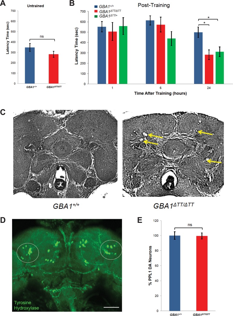
Fig 3. GBA1ΔTT homozygotes display a memory deficit and neurodegeneration, but do not have dopaminergic neuron loss.
(A) Latency time to initiate courtship in untrained 14-day-old males of indicated genotype. (B) Latency time to initiate courtship in 14-day-old males of indicated genotypes at 1 hour, 6 hours and 24 hours following training using a conditioned mating assay. Note the longer latency times in trained males (B) relative to untrained males (A). (C) Representative paraffin-embedded H&E-stained brain sections from 30-day-old controls and GBA1ΔTT homozygotes. Yellow arrows indicate vacuoles. (D) Representative image of a projected Z-series of a control adult Drosophila brain stained with anti-Tyrosine Hydroxylase to label dopaminergic (DA) neurons. DA neurons within the PPL1 cluster are indicated by the circled regions. Scale bar, 200 μm. (E) Relative number of DA neurons within the PPL1 cluster of 30-day-old GBA1ΔTT homozygotes (GBA1ΔTT/ΔTT) N = 19, normalized to age-matched WT controls (GBA1+/+) N = 21. There was no significant difference between the number of DA neurons within the PPL1 cluster per genotype by Student t test. Error bars represent s.e.m., ns indicates p>0.05, *p<0.05, **p<0.005 by Student t test in all results shown in this figure.

