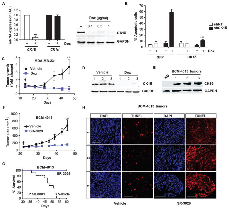
Fig. 3. Silencing or inhibition of CK1δ provokes breast tumor regression and blocks growth of PDX breast models.
(A) Left, qPCR data confirming efficient knockdown of CK1δ, but not CK1ε after 72 hours of treatment of MDA-MB-231 shCK1δ-expressing cells with Dox (0.3 μg/ml) (n=3; ***, p=0.0003). Right, corresponding CK1δ protein expression. (B) Cells expressing Dox-inducible CK1δ or non-targeting (NT) shRNA were treated with 1 μg/ml Dox for 72 hours and transfected with vectors expressing CK1δ or GFP cDNA resistant to shRNA. After a further 72 hours, percent cell death was measured by trypan blue dye exclusion in MDA-MB-231-shCK1δ (black) and cells expressing non-targeting shRNAs (NT, white) (n=4; ***, p=0.0001). (C) Growth of orthotopic MDA-MB-231-shCK1δ tumors in mice +/− Dox (administered ad lib in chow, 200 mg/kg), as monitored by caliper measurements (n=8 for each cohort; **, p=0.006). Arrow indicates addition of Dox. (D) CK1δ knockdown in tumor tissue isolated from three independent mice, 7 days after Dox administration began. (E) Immunoblot comparing CK1δ expression in extracts of normal human breast or three independent BMC-4013 PDX tumors. (F) Growth curves of BCM-4013 PDX tumors in mice treated with vehicle (black line) or SR-3029 (blue line). Arrow indicates timing of first dose (n=12 for each cohort; ***, p=0.0002). (G) Kaplan-Meier survival curve corresponding to studies shown in (F) (p value calculated using log-rank test). (H) TUNEL staining on serial sections of vehicle and SR-3029 treated BMC-4013 tumors (representative images are shown) (scale bar=200 μm).

