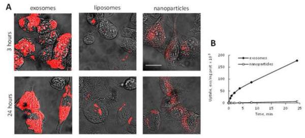
Figure 2. A profound accumulation of exosomes in 3LL-M27 cells in vitro.
3LL-M27 cells were incubated with fluorescently-labeled (red) exosomes, or liposomes, or PS NPs for various times and the amount of accumulated nanocarriers was examined by confocal microscopy (A), and spectrophotometry (B). Bar: 10 μm.

