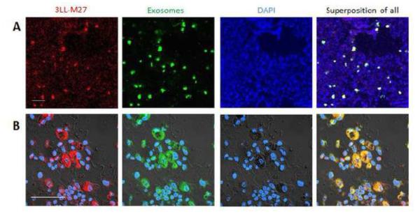
Figure 4. Co-localization of airway-delivered exosomes with pulmonary metastases.
Exosomes were isolated from macrophages conditioned media, and labeled with fluorescent dye, DID (green). C57BL/6 mice were i.v. injected with 3LL-M27 cells transduced with lentiviral vectors encoding the optical reporter mCherry (8FlmC) fluorescent protein. 21 days later, the mice with established pulmonary metastases (red) were i.n. injected with DID-labeled exosomes (green). 4 hours later, mice were euthanized, perfused, lungs were sectioned, and stained with DAPI (blue). The confocal images revealed near complete co-localization of exosomes with metastases (yellow). Images were obtained with ×10 (A), and ×60 (B) magnification. Bar: 50 μm.

