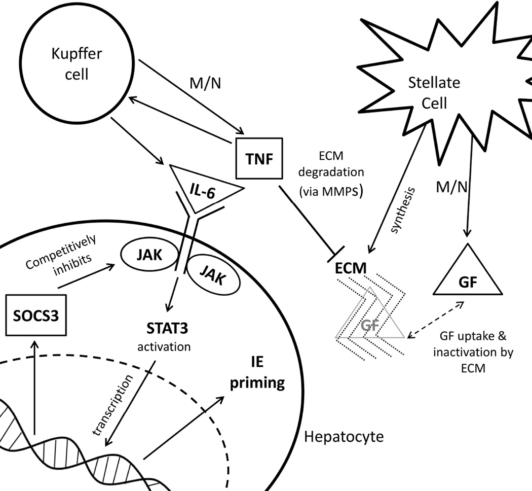
Figure 1. Biochemistry of liver regeneration for the model.
After partial hepatectomy, hepatocyte number is reduced which increases metabolic load (M/N). Increased metabolic load drives hepatocyte growth factor (GF) and tumor necrosis factor (TNF) expression. TNF degrades the ECM through matrix metalloproteases (MMPs). TNF also leads to the expression of IL-6, which initiates Janus kinase (JAK) signaling by activating STAT3. STAT3 leads to transcription of suppressor of cytokine signaling 3 (SOCS3), and intermediate-early genes (IE) that cause the hepatocyte to shift into a primed state. As regeneration progresses and hepatocyte numbers increase, TNF levels decrease and the ECM reforms and takes up GF. This stops the primed to replicating cell transition, and drives the replicating to quiescence cell transition.

