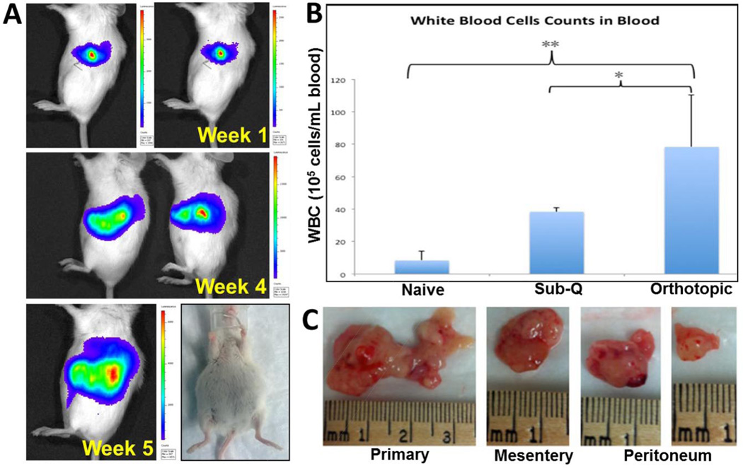
Figure 6.
Characterization of orthotopic pancreatic adenocarcinoma model in SCID beige mouse (A) Images showing the bioluminescence intensity obtained from IVIS imaging of mice injected with Panc-1 luc surgically in the pancreas with time post-surgery. All mice show accumulation of ascites fluid in their peritoneal cavity. (B) White blood cell count obtained from the blood of naïve, subcutaneous tumor and orthotopic tumor bearing mice, which is significantly higher for the tumor bearing animals; **(p < 0.01); * (p < 0.05)). (C) Image of primary tumors excised from the site of injection and secondary tumor nodules excised from mesentery and peritoneum.

