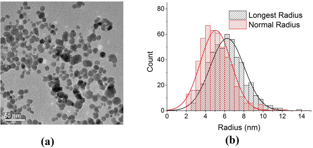
Fig 3.
(a) Room temperature transmission electron microscopy (TEM) images of aqueous EMG-308 IONPs were acquired with a FEI Tecnai T12 microscope (FEI, Inc., Hillsboro, OR) operating at 120 kV. A 200 mesh copper grid with formvar and carbon supports was dipped into a ~1 mg Fe/ml IONP suspension, then removed and allowed to dry before imaging. (b) The histogram of the longest radius and the radius normal to it were measured using Image J (NIH).

