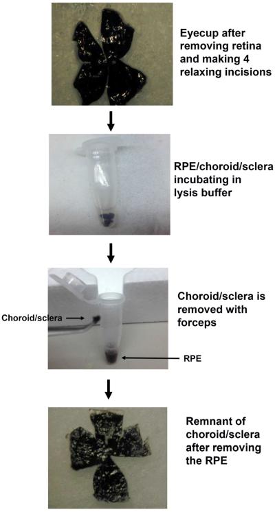
Figure 1.
Schematic diagram of isolated RPE using the new protocol. With the posterior eye cup, the retina is removed, and the RPE/Bruch's membrane/choroid/sclera is cut into four parts, and then incubated in protein lysis buffer. After gently tapping on the microcentrifuge tube, the RPE is separated from Bruch's membrane/choroid/sclera.

