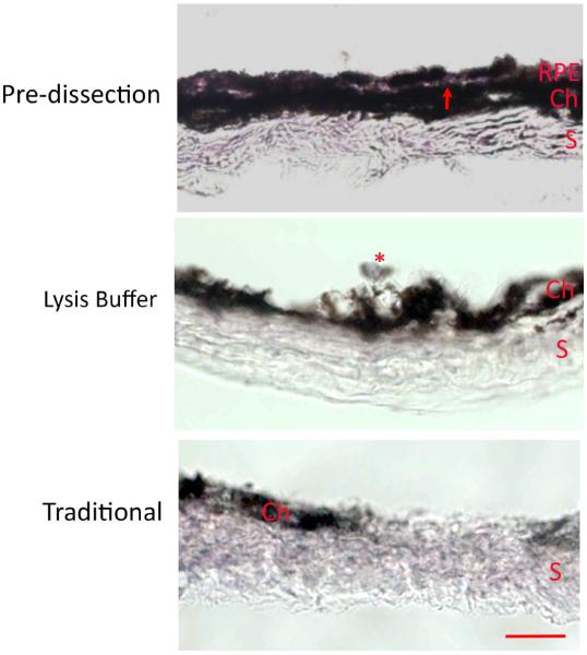
Figure 3.
Histological assessment of a C57BL/6J mouse eyes after lysis buffer and traditional dissection. Hematoxylin and eosin staining of the RPE/Bruch's membrane/choroid/scleral eyecup after the retina was removed. The eyecup pre-dissection shows an intact RPE layer. Note the thin clear line of Bruch's membrane (red arrow) that separates the choroid (Ch) from the RPE. The sclera (S) is also intact. After lysis buffer digestion, the RPE is gone, except for possibly a remnant of an RPE cell (*), with remaining choroid and sclera. After the traditional dissection, the sclera remains, with a remnant of the choroid (Ch). Bar=25 um.

