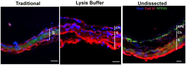
Figure 4.
Confocal fluorescence immunohistochemistry of C57BL/6x129 mouse eyecup for RPE65 and collagen VI. The eyecup after the traditional dissection of the RPE/choroid shows nonspecific immunolabeling for RPE65 (green) and collagen VI (red) in the sclera (S) only. The eyecup after lysis buffer digestion has collagen VI immunolabeling present in the choroid (Ch) and sclera (S), but no RPE65 immunolabeling. The undissected eyecup shows prominent RPE65 immunolabeling (green) in the RPE and collagen VI labeling in the choroid. Nonspecific labeling for RPE65 and collagen VI is seen in the sclera. Nuclei are labeled with Dapi (blue). Bar=15um.

