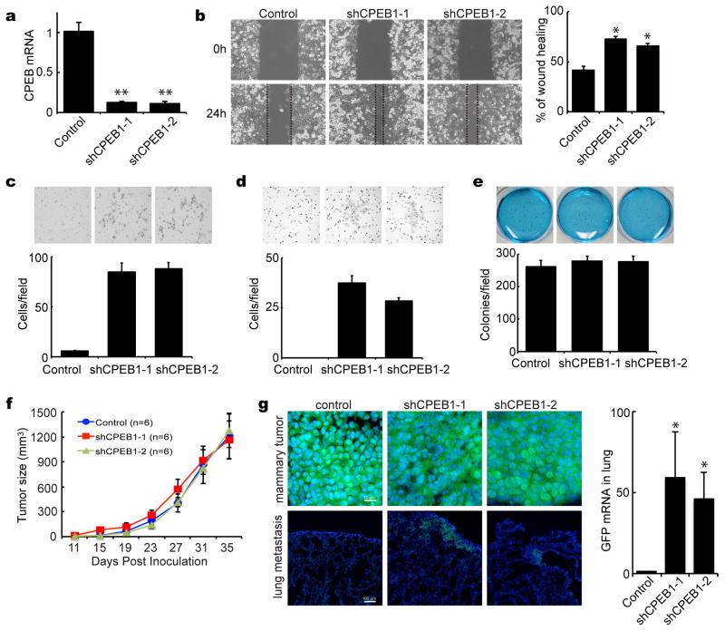
Figure 4.
CPEB1 depletion stimulates mammary tumor metastasis. (a) MCF7 cells were infected with lentiviruses expressing GFP only or GFP plus two different shRNAs against CPEB1. Real-time RT-PCR was used to assess CPEB1 depletion. (b) Wound-healing scratch in MCF-7 cells, some of which express CPEB1 shRNA. Migration assay (c), invasion assay (d) and soft-agar colony formation assay (e) following CPEB1 knockdown. (f) MCF7 cells infected with control or CPEB1 knockdown virus were inoculated into the fat pads of 8 week old female nude (nu/nu) mice. Calipers were used to measure tumor sizes along the two main axes. Tumor volumes were estimated by the relationship of the long axis × (short axis)2 × 0.5. (g) Metastatic MCF-7 cells in the lung were detected by immuno-fluorescence for GFP in tissue that was snap frozen and sectioned. Quantification of metastatic cells in the lung was determined by real-time RT-PCR for GFP.

