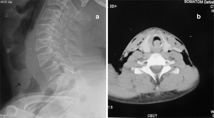
Fig. 1.
a X-ray soft tissue neck showing mass attached to anterior wall (marked with black arrow) in the cervical trachea just below the cricoid cartilage. b CT scan of neck revealed a mass attached to the anterior tracheal wall just below the cricoids cartilage, involving almost the entire tracheal lumen

