
FIG 2.
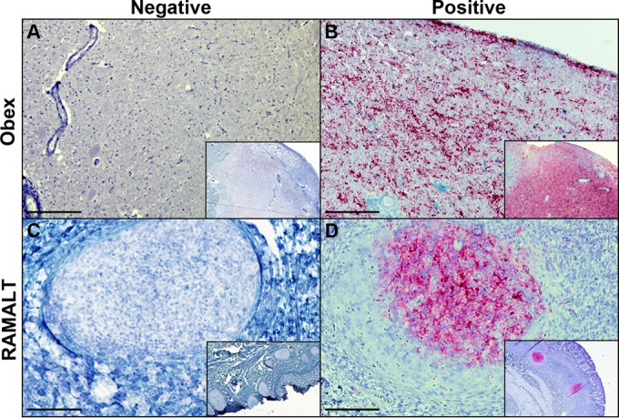
IHC detection of PrPCWD in brainstem (obex) and RAMALT samples by previously described protocols. Panels: A, CWD-negative obex section of an elk from RMNP; B, obex section of an elk from RMNP showing heavy accumulation of material staining positive for PrPCWD; C, RAMALT biopsy specimen from an elk from RMNP showing negative staining for PrPCWD; D, CWD-positive RAMALT biopsy specimen from an elk from RMNP showing heavy accumulation of material staining positive for PrPCWD. IHC analysis was performed with anti-prion 99 antibody (Ventana Medical Systems, Tucson, AZ). Bars = 250 μm.