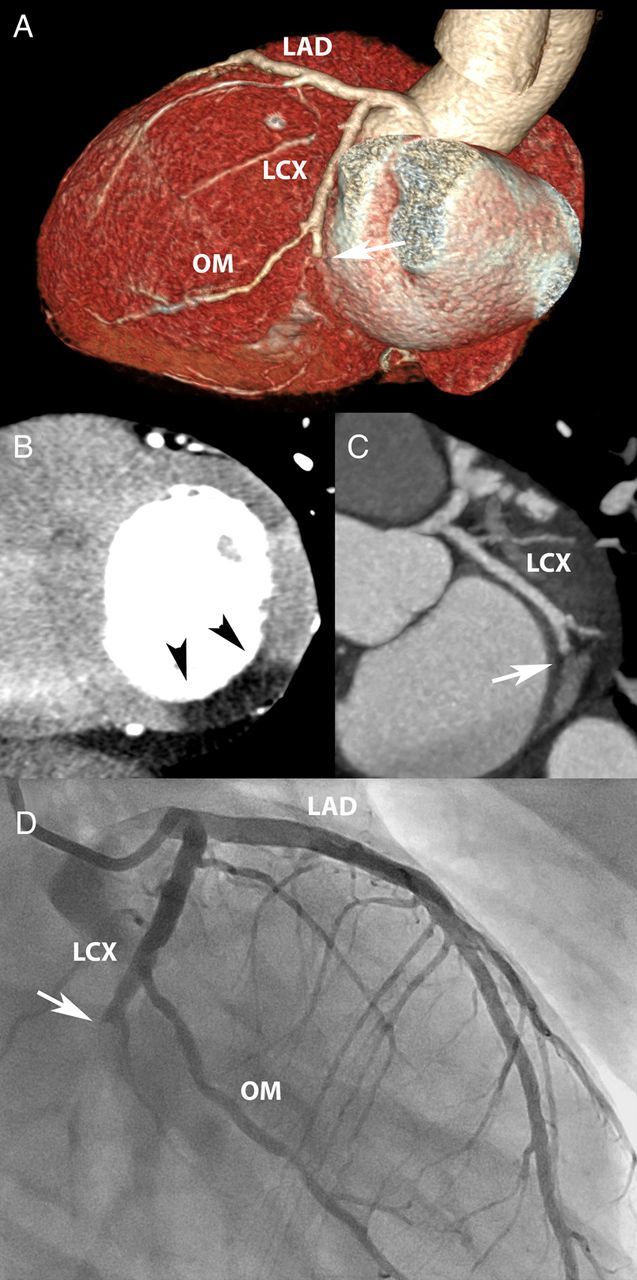
Figure 2.

Myocardial infarction. A 48-year-old man with position dependent, burning chest pain, and gastro-intestinal complaints. The conventional 12-lead electrocardiogram showed non-specific STT-segment changes. The conventional troponin T was borderline abnormal (0.04 mg/L). Coronary CTA (A and B) revealed an occluded distal LCX (arrow), with transmural hypo-enhancement and normal thickness of the infero-lateral wall (C, arrow heads). A subsequent rise in cardiac biomarkers (max CK-MB 93 µg/L) indicated acute myocardial injury, and invasive angiography confirmed an LCX occlusion (D, arrow).
