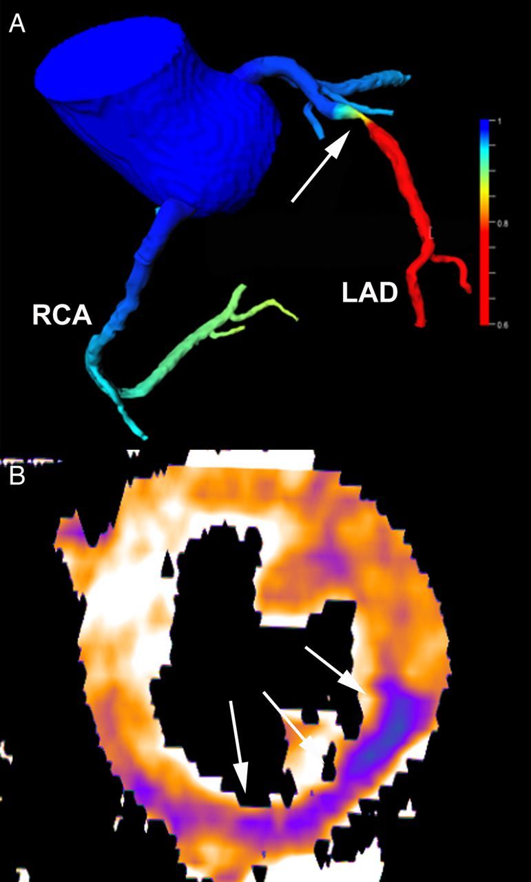
Figure 3.

Assessment of haemodynamic relevance by computed tomography. Computed tomography angiography-derived fractional flow reserve (A) shows a haemodynamic significant (red colour) stenosis in the LAD (arrow) with a simulated FFR value of 0.72. Stress perfusion computed tomography (B) demonstrating low myocardial blood flow (during pharmacological hyperaemia) in the infero-lateral wall (arrows) caused by an RCA stenosis.
