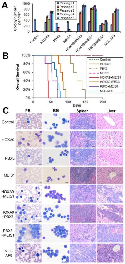
Figure 1. Co-expression of PBX3 and MEIS1 can transform normal mouse bone marrow (BM) progenitor cells in vitro and induce rapid AML in vivo.
(A) In vitro colony-forming/replating assays. Briefly, mouse normal BM progenitor (lineage negative; Lin-) cells collected from 4- to 6-week-old B6.SJL (CD45.1) mice were retrovirally co-transduced with MSCVneo+MSCV-PIG (Control), MSCVneo-HOXA9+MSCV-PIG (HOXA9), MSCVneo+MSCV-PIG-MEIS1 (MEIS1), MSCVneo+MSCV-PIG-PBX3 (PBX3), MSCVneo-HOXA9+MSCV-PIG-MEIS1 (HOXA9+MEIS1), MSCVneo-HOXA9+MSCV-PIG-PBX3 (HOXA9+PBX3), MSCVneo-MEIS1+MSCV-PIG-PBX3 (PBX3+MEIS1), or MSCVneo-MLL-AF9+MSCV-PIG (MLL-AF9). The colony cells were replated every 7 days for up to 5 passages and colony numbers were counted for each passage. Mean±SD values of colony counts are shown. (B) Mouse BM transplantation (BMT) assays were conducted for the above 8 groups with the first-passage colony cells (CD45.1) as donors, which were transplanted into lethally irradiated 8- to 10-week-old C57BL/6 (CD45.2) recipient mice. Kaplan-Meier curves are shown. Five mice were studied in each group, except for the MLL-AF9 group in which 6 mice were studied. (C) Cell/tissue morphologies of the 8 groups. Peripheral blood (PB) and BM cells were stained with Wright-Giemsa. The spleen and liver tissues were stained with hematoxylin and eosin (H&E). The length of bars represents 10 μm for PB and BM, and 100 μm for spleen and liver.

