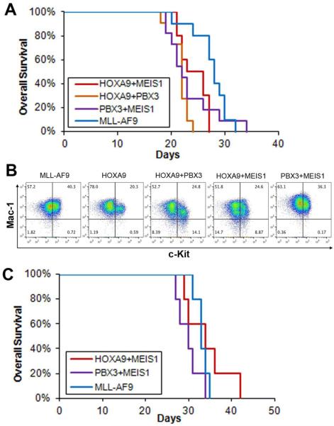
Figure 2. PBX3/MEIS1-induced AML is transmissible in secondary transplantation recipients.
(A) Kaplan-Meier survival curves of secondary transplantation recipient (CD45.2+) mice transplanted with primary leukemic BM cells (CD45.1+) of the HOXA9+MEIS1 (recipient mouse number: n=10), HOXA9+PBX3 (n=11), PBX3+MEIS1 (n=11) and MLL-AF9 (n=10) groups. Primary AML BM cells from two donor mice were used for each group. There is no significant difference (p>0.1) between survival of the PBX3+MEIS1 group and that of any other three groups. (B) Flow cytometry analysis of leukemic BM cells from the above secondary BMT recipient mice. Antibodies against Mac-1 and c-Kit were used. Flow data of leukemic BM samples from one recipient mouse is shown as representative for each group. (C) Kaplan-Meier survival curves of secondary transplantation recipient (CD45.2+) mice transplanted with primary leukemic spleen cells (CD45.1+) of the HOXA9+MEIS1 (n=5), PBX3+MEIS1 (n=5) and MLL-AF9 (n=5) groups. Primary AML spleen cells from one donor mouse were used for each group. There is no significant difference (p>0.1) between survival of the PBX3+MEIS1 group and that of any other two groups.

