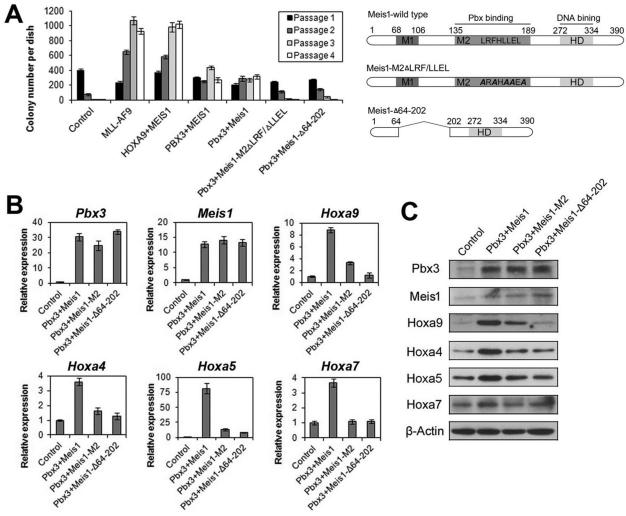
Figure 6. The binding between Meis1 and Pbx3 is critical for their synergistic effects on cell transformation and up-regulation of homeobox genes.
(A) In vitro colony-forming/replating assays. Briefly, mouse normal BM progenitor (lineage negative; Lin-) cells collected from 4- to 6-week-old B6.SJL (CD45.1) mice were retrovirally co-transduced with MSCVneo+MSCV-PIG (Control), MSCVneo-MLL-AF9+MSCV-PIG (MLL-AF9), MSCVneo-HOXA9+MSCV-PIG-MEIS1 (HOXA9+MEIS1), MSCVneo-MEIS1+MSCV-PIG-PBX3 (PBX3+MEIS1), MSCVneo-Meis1+MSCV-PIG-Pbx3 (Pbx3+Meis1), MSCVneo-Meis1-M2ΔLRF/ΔLLEL+MSCV-PIG-Pbx3 (Pbx3+Meis1-M2ΔLRF/ΔLLEL) or MSCVneo-Meis1-Δ64-202+MSCV-PIG-Pbx3 (Pbx3+ Meis1-Δ64-202). The colony cells were replated every 7 days for up to 4 passages and colony numbers were counted for each passage. Mean±SD values of colony counts are shown (left panel). The domain structure of the wild-type and mutant Meis1 proteins are also shown (right panel). (B,C) qPCR (B) and Western blotting (C) analyses of expression levels of a series of representative Homeobox genes in colony cells from the first passage of four groups (samples generated from colony-forming/replating assays shown in Figure 6A). Mean±SD values are shown in Figure 6B.

